
who we are
Calgary Vision Centre is a full scope Optometry clinic located in downtown Calgary. The first set of eyes we examined was in 1998, and over a quarter million eyes later we are proud to still be locally owned and operated, allowing us to continue to care for the next quarter million eyes in the best way we see fit.

our doctors

Dr. Robert Burke: Dr. Burke was educated in Chicago, IL, and has spent time training in various settings, including the Vision Institute of Canada, the Illinois Eye Institute and the Newington VA Hospital, but for the last 17 years he has practiced exclusively at Calgary Vision Centre. His passion for advanced technology is what defines Calgary Vision Centre's philosophy. His examinations are meticulously performed and he always reserves time to carefully explain his findings and answer all client questions. He is constantly exploring novel ways of applying advanced technology to the eye health world. He is the inventor of a suite of novel eye charts that fuse the legacy eye charts that have been used for over a century (the “Big E” chart) rendered over top of real-world 4k video, in an effort to replicate real world vision demands. He is also the creator of a successful iOS app (Parks 3 Step) that aids fellow optometrists in accurately diagnosing specific eye conditions. Recently, he has been concentrating his efforts on leveraging VR headsets and gaze tracking software to explore the potential correlation between eye teaming inaccuracies and visual discomfort (eyestrain, headaches, light sensitivity) particularly in patients recovering from concussions.

Dr. Justin Norris: Dr. Norris started his practice at Calgary Vision Centre (formerly Gulf Square Optometry) in 1998 and has learned over the years that each patient comes with a unique set of visual needs and expectations. He understands that even the smallest change in prescription, the specific brand of lens chosen, or a subtle variation in lens fit can make or break the success of an eyewear solution. An eye exam with Dr. Norris lasts about thirty minutes, and most of that time is dedicated to discussing your individual options—in some cases, those discussions even lead to the recommendation of going without a prescription at all. In addition to his routine practice, Dr. Norris has considerable experience from his time working at a laser centre, and he has undergone laser surgery himself. This firsthand experience makes him exceptionally comfortable and knowledgeable when discussing advanced treatment options with his patients. Beyond his professional endeavors, he takes great pride in being an optometrist, while also valuing precious time spent with his family, and contributing to his community as a coach in hockey, soccer, and ringette.
transforming vision care with innovative optometry
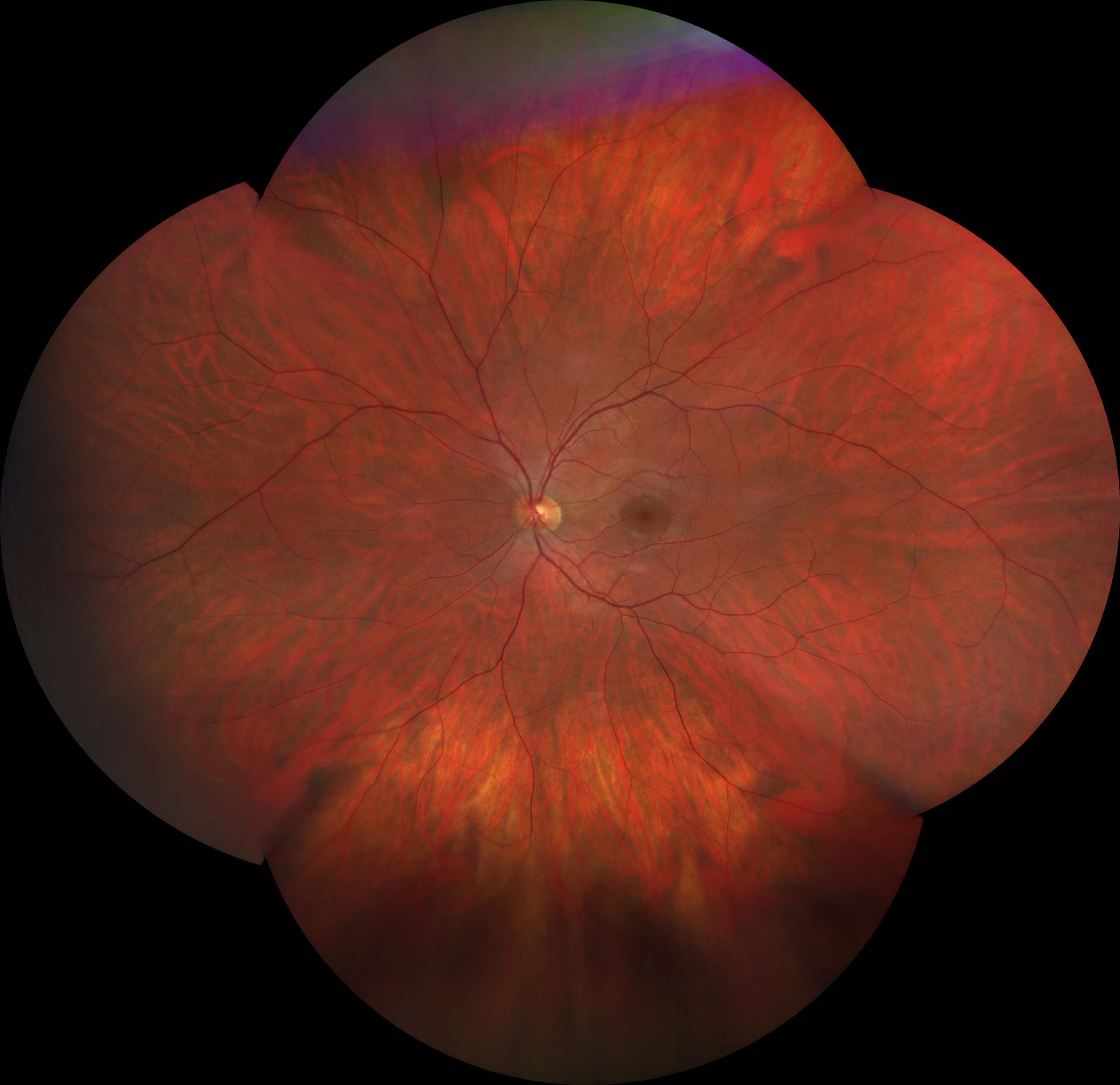
Figure 1: A patient’s retinal image captured at Calgary Vision Centre March 22, 2024.
The image above is an actual image captured in our clinic by our equipment and rendered in office.
The orange tint above isn’t mere art—it is the retina, a fragile, photoactive layer lining the interior of our eyes. Because light enters only through the narrow pupil, which constricts further in bright light, capturing a clear image of the retina is no simple task; the familiar “red eye” seen in flash photography merely confirms its presence.
For a thorough view, clinicians dilate the pupil—a gold standard, albeit time-consuming, procedure that carries some risks. Since 2004, our clinic has evolved through five distinct imaging eras, consistently pioneering new technology. This steadfast commitment to innovation has not only sharpened our diagnostic capabilities but also set a high standard for forward-thinking eye care
the analog era (pre - 2005)

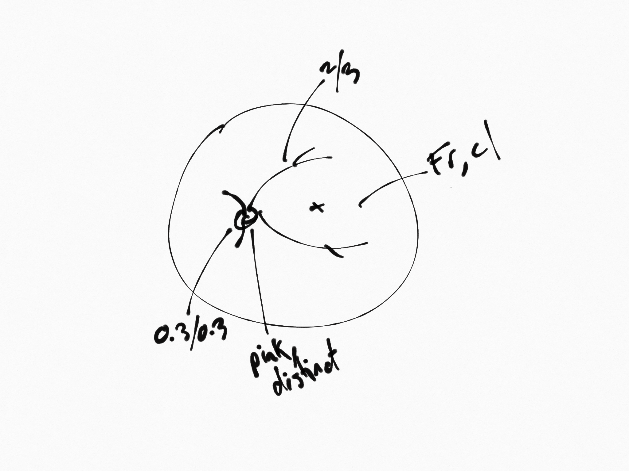
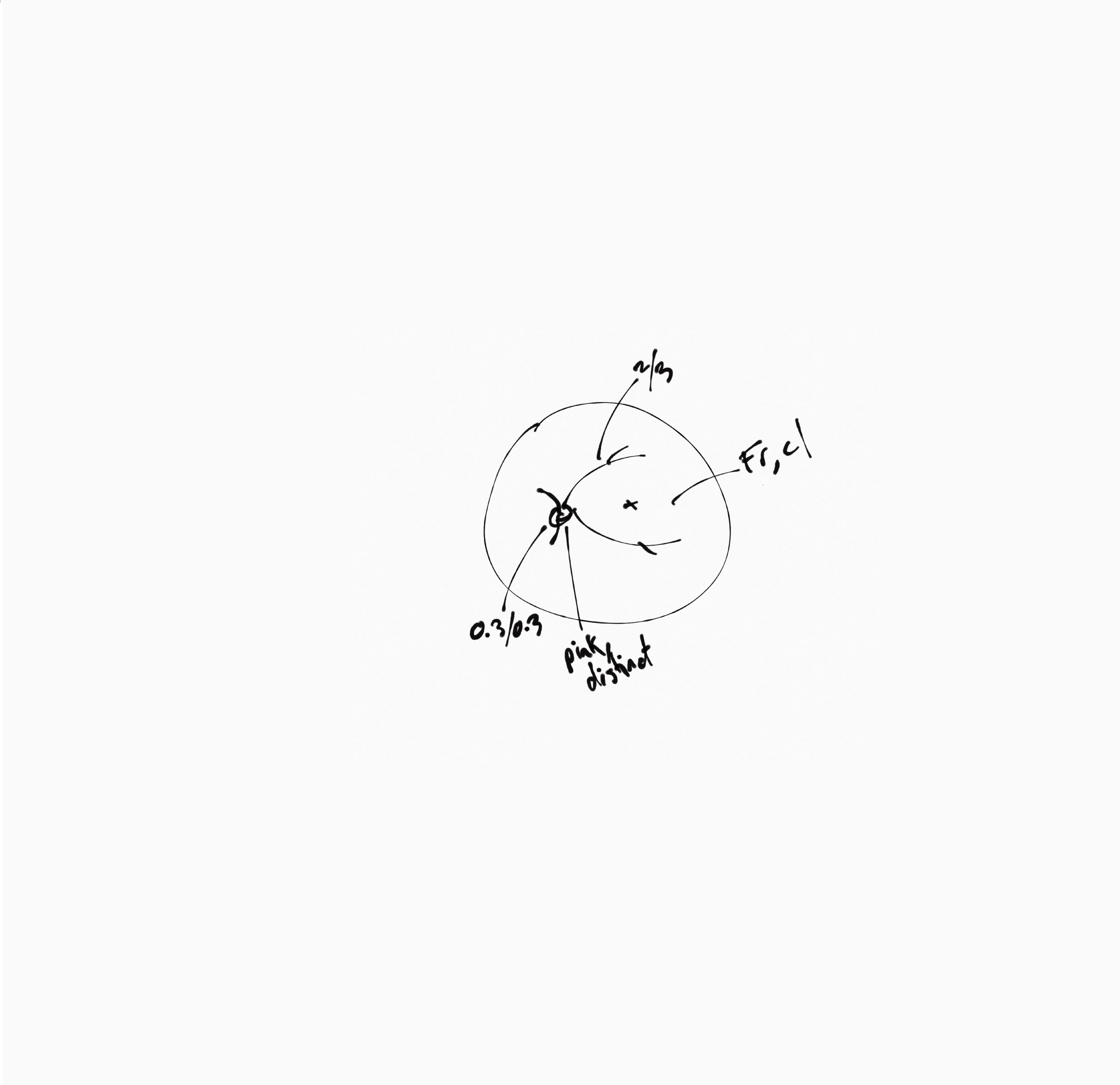
A handheld instrument—a classic ophthalmoscope—is directed into the eye, projecting a narrow beam that serves as a spotlight on the retina. The clinician must skillfully maneuver this beam across the retinal surface, effectively "painting" a larger picture of the tissue. Because the ophthalmoscope does not capture digital images, any noteworthy observations are traditionally recorded as simple, hand-drawn sketches. This is the very method your family doctor employs during a routine eye exam. Its affordability and portability have ensured that the handheld ophthalmoscope remains a cherished tool in clinics worldwide. The illustration on the right, for instance, depicts the results of an undilated ophthalmoscopic examination performed on our patient from Figure 1—revealing sketches that, while not technically inaccurate, display a surprising lack of detail.
the digital era (2005 - 2011)
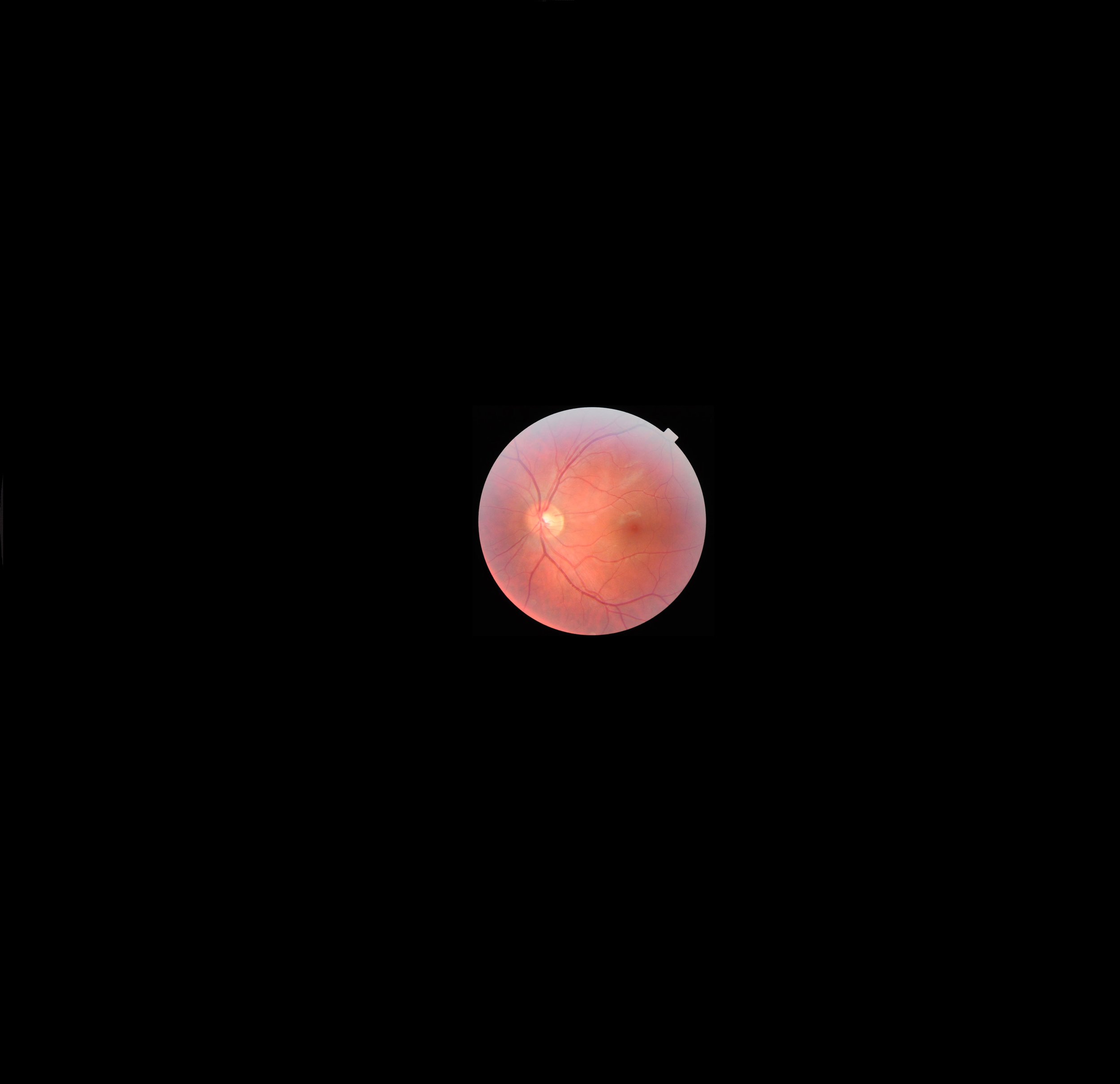
The acquisition of a color retinal camera marked a transformative advance for our practice—enhancing both diagnostic capabilities and the precision of our record keeping. With the ability to capture a single, detailed color image of critical regions of the retina, we gained the power to examine this delicate tissue in unprecedented detail and to reference these images over time—a development that was nothing short of revolutionary at the time.
In the image presented, the retinal tissue begins to mirror that seen in Figure 1, with a familiar network of blood vessels and anatomical structures that a trained clinician can easily recognize. However, the field of view in this photograph is relatively narrow, covering only about a 30° segment of the retina. The surrounding darkness represents additional areas that this technology cannot yet capture. Notably, the yellow circle at the center demarcates the optic nerve; when compared with Figure 1, it becomes apparent just how zoomed in this particular image is.
the 3D era (2011 - present)
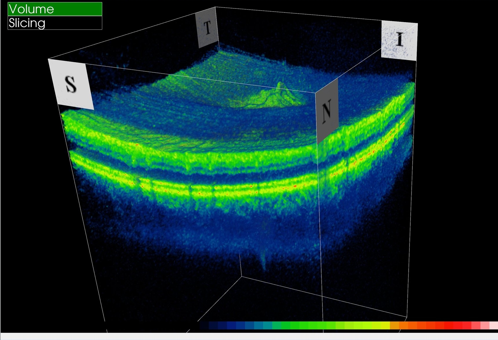
Currently, our imaging remains firmly rooted in the 3D era, yet we have upgraded our OCT scanner twice since 2011—each enhancement delivering a dramatic leap in resolution. Although this technology has not expanded our field of view beyond the 30° limit, its true strength lies in its ability to capture subsurface details, including depth and elevation data within that narrow scope. Moreover, the system automatically measures and tracks the size of key retinal structures, cross-referencing these values against a normative database to promptly flag any potential concerns.
The ultra-wide field era (2018 - present)

With the advent of the 3D era significantly enhancing our ability to diagnose and monitor minute changes in localized regions of the retina, a new chapter has begun—the ultra-wide field era. This innovation extends our vision beyond the conventional 30° zone, allowing us to explore previously inaccessible areas of the retina. In the image above, one can observe the same retinal area both before and after the deployment of the Zeiss Clarus 500 ultra-wide field camera. It is striking how much more of the retina this camera reveals compared to its predecessors, all while preserving true-to-life color—a vital attribute when identifying subtle, freckle-like lesions that might indicate pathology. Moreover, this camera leverages a feature called 'fundus auto-fluorescence' to detect areas of imminent cell death, marking a significant leap in our diagnostic capabilities.
The image featured in Figure 1 is an ultra-wide field montage obtained with the Zeiss Clarus 500. This comprehensive view is further enriched by integrating data from the Topcon 3D OCT, all rendered seamlessly in-office to provide a multidimensional perspective on retinal health.
the virtual reality era (2022 - present)
Our transformative journey through the realm of retinal imaging and diagnostic precision has led us to an extraordinary new chapter in eye care. Since 2022, we have fully embraced the virtual reality revolution by incorporating two state-of-the-art VR headsets into our practice. These cutting-edge devices gather unparalleled data on peripheral vision and the intricate coordination of your eyes as they work in harmony. This revolutionary technology brings together decades of progress—from the era of manual ophthalmoscopy to advanced multi-modal imaging and expansive ultra-wide field photography. By integrating these VR-based insights with our traditional imaging methods, we deliver a seamlessly holistic, personalized assessment of your visual performance. This innovative approach, exclusive to our clinic, not only exemplifies our dedication to excellence but also sets a new benchmark for precision and customization in modern eye care.